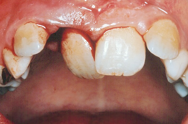
Lateral luxation


LUXATION IN A PALATAL DIRECTION (Lateral luxation)
* Administer local anesthesia.
* Palpate the sulcus fold, and localize the displaced root apex. Apply firm, digital pressure
in an incisal direction and move the tooth back through the fenestration into the socket.
* Reposition the tooth back to its original position by axial pressure.
* Reposition fractured bone with finger pressure.
* Take a radiograph to verify correct position.
* Stabilize the tooth with a splint.
* Maintain the splint for a minimum of 3-4 weeks.
* Take a radiograph after about 3 weeks. With signs of marginal bone breakdown, the
splint is maintained for another 3-4 weeks.
Follow-up:
All traumatized teeth are carefully observed for clinical and radiographic signs of complications. The intervals between
re-examinations should be individualised depending upon the severity of traume, the expected type of complication and the age
of the patient. When external inflammatory root resorption is an expected complication, a clinical and radiographic control
should be taken every 14 days until the situation is clear. In lateral luxation the possibility of external inflammatory root
resorption is rather low, below 5 %.
Endodontic considerations:
In mature teeth where the apex is closed, lateral luxation often results in pulpal necrosis. If pulpal necrosis develops,
endodontic therapy is performed.