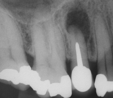

The area was anaesthetised and a marginal flap was opened. The lesion, which had perforated through the buccal cortical bone, was removed with hand instruments. The root tip was cut off with a bur and the apical root filling was removed using small ultrasound tips. The canal was them prepared by diamond coated ultrasound tips and bend Hedstroem hand files. Physiological saline was constantly used for irrigation of the area and the tooth. A radiograph and an operating microscope verified a clean root canal apically to the post.