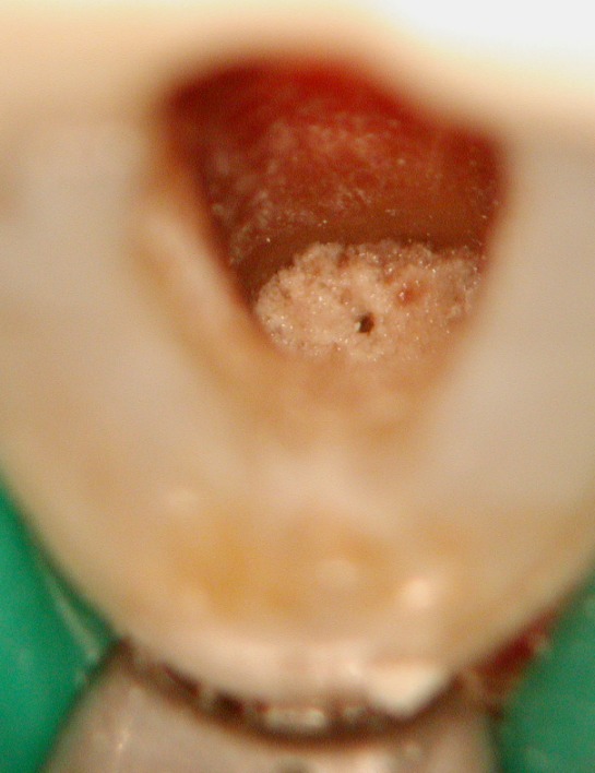
The Rubber dam was placed, working field disinfected, and the temporary filling removed. The canal was examined by using an
operating microscope to evaluate possibilities to locate the original root canal. Instead of "normal" secondary dentin, the canal
space was filled with osteoid like tissue.
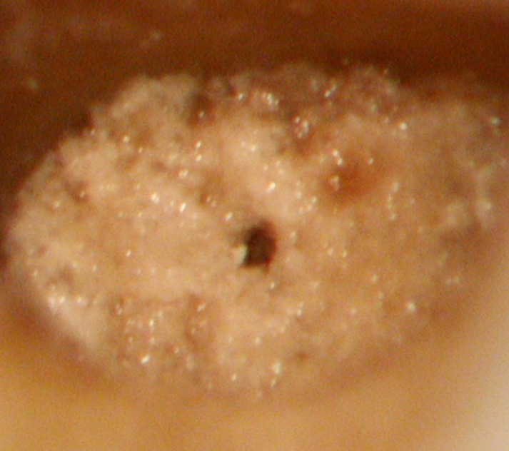
Higher magnification
revealed porosities in the hard tissue with obvious remnants of necrotic tissue.