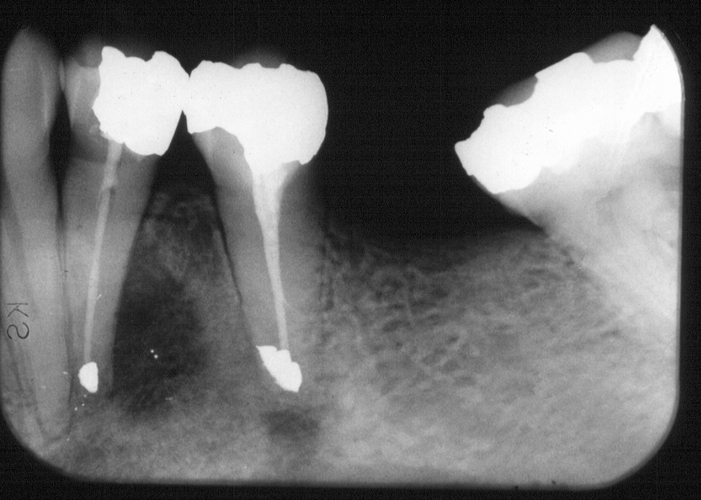
A control radiograph taken one year later showed considerable healing: only small changes could be seen at the apical area of tooth 35 and the bone cavity distally to 34 had reduced to one fourth of the size of the original lesion. The teeth had been symptomfree after the treatment.