D16


#16. Maxillary
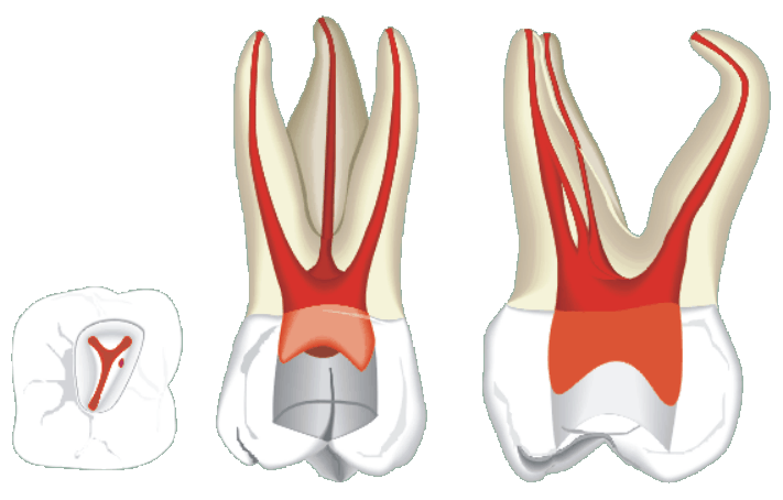
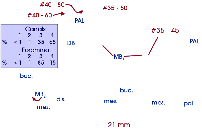
molars have from one to three roots and from two to four root canals. From
an occlusal view the pulp chamber is situated rather mesially, which has to
be taken into account when cutting the access cavity.
The upper first molar is perhaps the most variable tooth when it comes to
root canal morphology, and provides quite a challenge in endodontics. There
are usually three roots with three or four root canals. Dentists are quite
familiar with the mesiobuccal, distobuccal and palatal canals, but not with
the fourth canal, which is known as the mesiocentric or mesiopalatal, mb2
or accessory mesiobuccal canal. This fourth canal is usually difficult to
find just by clinical inspection and is not apparent in the radiograph. However,
finding all canals is necessary for successful therapy.
The distobuccal canal is often easy to locate and instrument. It is typically
rather straight or curves only slightly mesially, or sometimes distally.
The palatal canal always looks straight radiographically but often has a buccal
curvature. If this curvature is not identified by careful exploration with
files it can lead to perforation 2 - 4 mm before the apex. Moreover, in radiographs
a file will still appear to be in the canal but in reality it is only superimposed
onto the canal. The palatal canal is often 1 - 2 mm longer than the buccal
canals. Two palatal roots in the upper first molar have been reported in the
literature.
The mesiobuccal root is the most challenging to treat. The root is usually
curved all the way to the apex, which increases the risk of tip perforation
and strip perforation. The distal surface of the root is concave which increases
the risk of strip perforation.
The mesiopalatal canal is present in well over half of cases, with some authors
reporting over 90% incidence. The canal orifice is difficult to find because
it is typically situated near the mesial wall of the pulp chamber. While the
other three canals can readily be found, the fourth canal must always be actively
looked for with suitable instruments. The orifice is usually located 1 - 3
mm palatally from the mesiobuccal canal. In most cases the mesiopalatal canal
joins the mesiobuccal canal before the apex.