D46


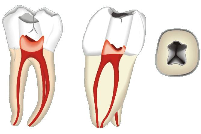
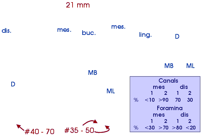
#46. The mandibular first molar is perhaps the most frequently endodontically treated molar. It is, however, often quite difficult to treat because of its root canal anatomy. It usually has 3 - 4 canals, two in the mesial root and one or two in the distal root. The Distal canal(s) is normally straight all the way to the apex, oval or flattened in cross-section, but quite large, which makes instrumentation easy. Often the most apical 1 - 2 mm of this canal curves up to 90 degrees distally, but this is seldom a clinical problem. The distal canal may also curve mesially, but the curvature is not sharp and usually remains easy to instrument. The mesial canals in the first molar are often a challenge for the dentist. Both the mesiobuccal and mesiolingual canals are usually curved along their whole length, and the curvature is typically greatest in the apical region. The canals curve distally, but they also curve buccally or lingually at the same time. Bucco-lingual curvatures are not readily seen in the radiograph, which emphasizes the importance of the dentist's knowledge of possible variations in canal morphology. One must routinely search for four canals in the lower first molar. The distal canals often start together and separate a few millimeters below the pulp chamber floor. Both distal and mesial canals can join before the apex. This is important to detect before obturation, to gain optimal results. Mandibular first molars with two canals are rare. Usually, finding only two canals indicates that the mesiobuccal canal has not yet been located.