Perforations: Treatment

The treatment
and prognosis of perforations is largely dependent on the following factors:
1) presence of bacterial infection, 2) level of perforation related to the
bone level, 3) the size of the perforation.
In infected perforations, the area must first be cleaned and disinfected e.g.
with calcium hydroxide treatment. In cases of bone destruction, clear signs
of healing should be observed before obturation. Small perforations to bone
can then be repaired with MTA, IRM, super-EBA, Cavit-R, or gutta-percha and
sealer. In perforations above the bone level the use of composite materials
should be considered. In larger perforations surgery is often needed to obtain
proper sealing. In such cases, composite materials and MTA should be considered
as materials of choice.
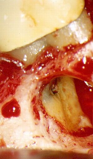
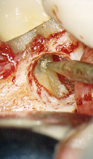
In non-infected perforations (e.g. treatment of pulpitis), the perforation
should be closed immediately if possible. Bleeding is first controlled by
irrigation with sterile physiological saline or saturated calcium hydroxide,
or by calcium hydroxide compression. Marginal dentine is then cleaned to facilitate
a good seal and the perforation is then filled with a material as suggested
above. Knowledge of the respective properties of each material is an essential
prerequisite for success. An operating microscope or magnification loupes
are excellent aides in the treatment of perforations.