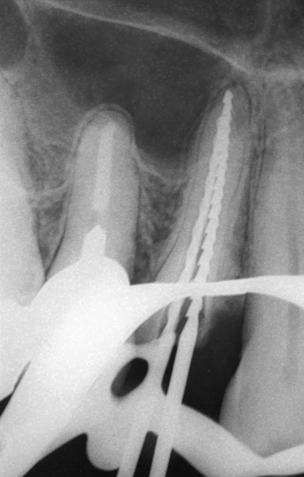
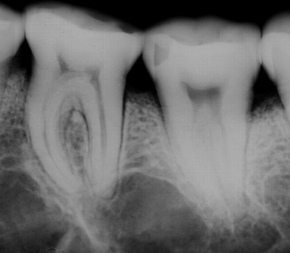
Anatomical findings - Radiolucent


Several anatomical features in radiographs can show some similarities to apical or lateral periodontitis. The most common are the maxillary sinus (upper canine - molar regions), mental foramen (lower premolars) and incisive foramen (upper centrals), variations in normal bone structure and nutritional canals. When in doubt, pulp sensibility testing and several x-ray projections to study the continuity of the apical PDL usually help in decision making.