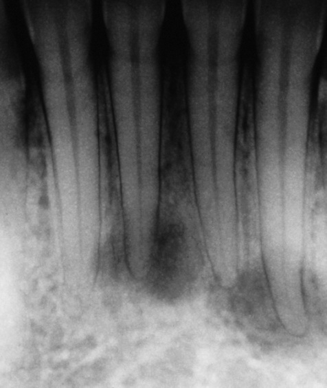
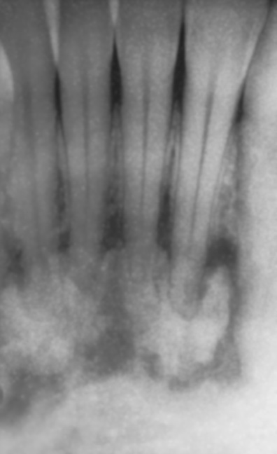
Periapical cemental dysplasia - Radiolucent


Periapical cemental dysplasia can be easily misdiagnosed as apical periodontitis, particularly in its early stage. In the initial phase, cellular fibrous tissue is replacing bone at the tooth apices. At a later stage, cementum-like tissue is produced, changing the radiolucent lesion into a more radiopaque one. Generally, such teeth are vital with no pulpal pathology. Periapical cemental dysplasia is of unknown aetiology, it is usually asymptomatic and does not require treatment. It is most common in young females and in the mandibular anterior region.